
Microbiology & Science

BACTERIA
Bacteria are microbes with a cell structure simpler than that of many other organisms. Their control center, containing the genetic information, is contained in a single loop of DNA. Some bacteria have an extra circle of genetic material called a plasmid rather than a nucleus. The plasmid often contains genes that give the bacterium some advantage over other bacteria. For example, it may contain a gene that makes the bacterium resistant to a certain antibiotic.
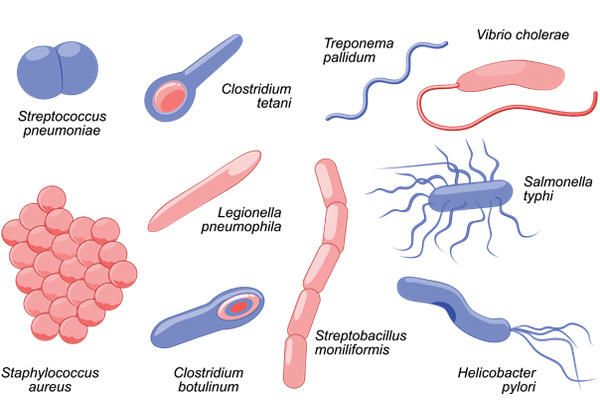
Bacteria are classified into five groups according to their basic shapes: spherical (cocci), rod (bacilli), spiral (spirilla), comma (vibrios) or corkscrew (spirochaetes). They can exist as single cells, in pairs, chains, or clusters.
Bacteria are found in every habitat on Earth: soil, rock, oceans and even arctic snow. Some live in or on other organisms including plants and animals including humans. There are approximately 10 times as many bacterial cells as human cells in the human body. A lot of these bacterial cells are found lining the digestive system. Some bacteria live in the soil or on dead plant matter where they play an important role in the cycling of nutrients. Some types cause food spoilage and crop damage but others are incredibly useful in the production of fermented foods such as yoghurt and soy sauce. Relatively few bacteria are parasites or pathogens that cause disease in animals and plants.

How do bacteria reproduce?
Most bacteria reproduce by binary fission. In this process, the bacterium, which is a single cell, divides into two identical daughter cells. Binary fission begins when the DNA of the bacterium divides into two (replicates). The bacterial cell then elongates and splits into two daughter cells each with identical DNA to the parent cell. Each daughter cell is a clone of the parent cell.
When conditions are favorable such as the right temperature and nutrients are available, some bacteria like Escherichia coli can divide every 20 minutes. This means that in just seven hours one bacterium can generate 2,097,152 bacteria. After one more hour, the number of bacteria will have risen to a colossal 16,777,216. That’s why we can quickly become ill when pathogenic microbes invade our bodies.
Survival mechanism
Some bacteria can form endospores. These are dormant structures, which are extremely resistant to hostile physical and chemical conditions such as heat, UV radiation and disinfectants. This makes destroying them very difficult. Many endospore-producing bacteria are nasty pathogens, for example Bacillus anthracis, the cause of anthrax.

Classification of Bacteria on the basis of Nutrition
- Nutrition is substances used in biosynthesis and energy production and therefore are required for all living things.
- Bacteria, like all living cells, require energy and nutrients to build proteins and structural membranes and drive biochemical processes.
- Bacteria require sources of carbon, nitrogen, phosphorous, iron and a large number of other molecules.
- Carbon, nitrogen, and water are used in the highest quantities.
- The nutritional requirements for bacteria can be grouped according to the carbon source and the energy source.
- Some types of bacteria must consume pre-formed organic molecules to obtain energy, while other bacteria can generate their own energy from inorganic sources.
Nutritional Types of Bacteria
On the basis of energy source organisms are designated as:
Phototrophs:
- The organisms which can utilize light as an energy source are known as phototrophs. These bacteria gain energy from light.
Chemotrophs:
- These bacteria gain energy from chemical compounds. They cannot carry out photosynthesis.
On the basis of electron source organisms are designated as:
Lithotrophs:
- Some organisms can use reduced organic compounds as electron donors and are termed as Lithotrophs.
- They can be Chemolithotrophs and Photolithotrophs
Organotrophs:
- Some organisms can use organic compounds as electron donors and are termed as organotrophs.
- Some can be Chemoorganotrophs and Photoorganotrophs.
Thus, bacteria may be either:
- Photo-lithotrops: These bacteria gain energy from light and use reduced inorganic compounds such as H2S as a source of electrons. eg: Chromatium okeinii.
- Photo-organotrophs: These bacteria gain energy from light an d use organic compounds such as Succinate as a source of electrons.eg; Rhodospirillum.
- Chemo-lithotrophs: These bacteria gain energy from reduced inorganic compounds such as NH3 as a source of electron eg; Nitrosomonas.
- Chemo-organotrophs: These bacteria gain energy from organic compounds such as glucose and ammino acids as a source of electrons.eg; Pseudomonas pseudoflora.
- Some bacteria can live ether chemo-lithotrophs or chemo-organotrophs like Pseudomonas pseudoflora as they can use either glucose or H2S as electron source.
On the basis of carbon source bacteria may be:
- All organisms require carbon in some form for use in synthesizing cell components.
- All organisms require at least a small amount of CO2.
- However, some can use CO2 as their major or even sole source of carbon; such organisms are termed as Autotrophs (Autotrophic bacteria).
- Others require organic compounds as their carbon source and are known as Heterotrophs (Heterotrophic bacteria).
Biochemical Test
Biochemical tests are the tests that are performed on different bacteria for their identification on the basis of their biochemical activities towards different biochemical compounds.
- Biochemical tests are one of the traditional methods for the identification of microorganisms, usually performed with phenotypic identification.
- For many years these methods were employed extensively, and they continue to be used nowadays, especially in some laboratory routines where a particular type of microorganism has to be identified rapidly.
- The ability of microorganisms to utilize certain biomolecules, resulting in useful organic compounds for themselves forms the basis of various biochemical tests.
- Biochemical tests are of different types, where the identification or distinction between different microorganisms is made on various bases.
- One of the traditional methods commonly used is a simple visual detection of the growth of the organism in the presence of essential nutrients by increased turbidity in the liquid medium.
- In other tests, however, the results are based on the change in color of the medium as a result of the change in the pH of the medium.
- Microorganisms can be classified into different groups on the basis of their reaction to such tests. Some tests even allow the distinction of microorganisms to the species level.
- Biochemical tests are thus, essential as they are inexpensive and relatively simple to perform.
- The physiology of bacteria and other microorganisms differs from one another, which allows for the differentiation of such microorganisms.
- Biochemical tests, however, have some disadvantages. Despite being inexpensive and allowing both quantitative and qualitative information about the diversity of microorganisms present in a sample, these methods are laborious and time-consuming, and results are only observed after several days.
- In some cases, false positives are obtained, especially when considering similar microbial species.
Pond bacteria
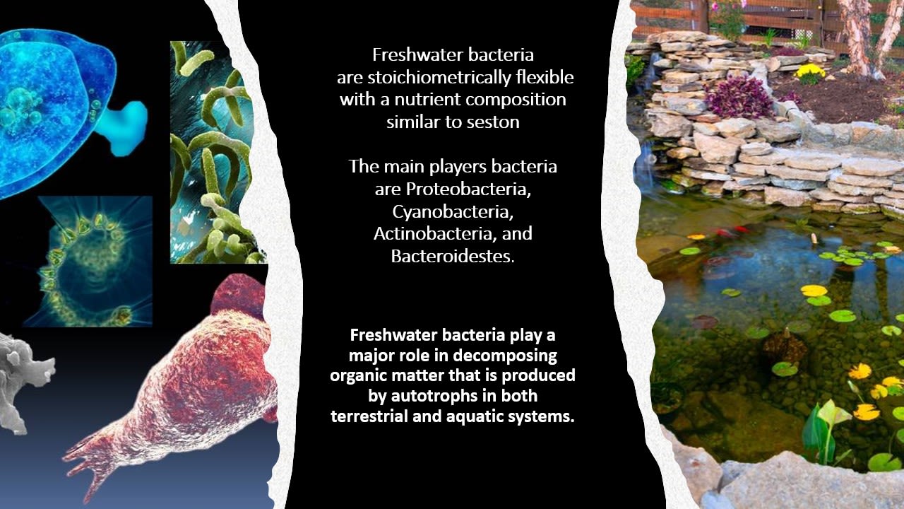

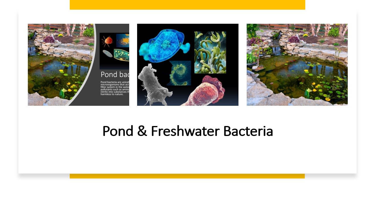
Pond & Fresh Bacteria

Stylonychia
Stylonychia is a genus of ciliates, in the subclass Hypotrichia. Species of Stylonychia are very common in fresh water and soil, and may be found on filamentous algae, surface films, and among particles of sediment.

Lepadella rotifer
Lepadella rotifer. Polarised light micrograph of a Lepadella sp. rotifer. Rotifers are microscopic aquatic animals that are related to roundworms. Magnification: x200 when printed 10 centimetres wide.
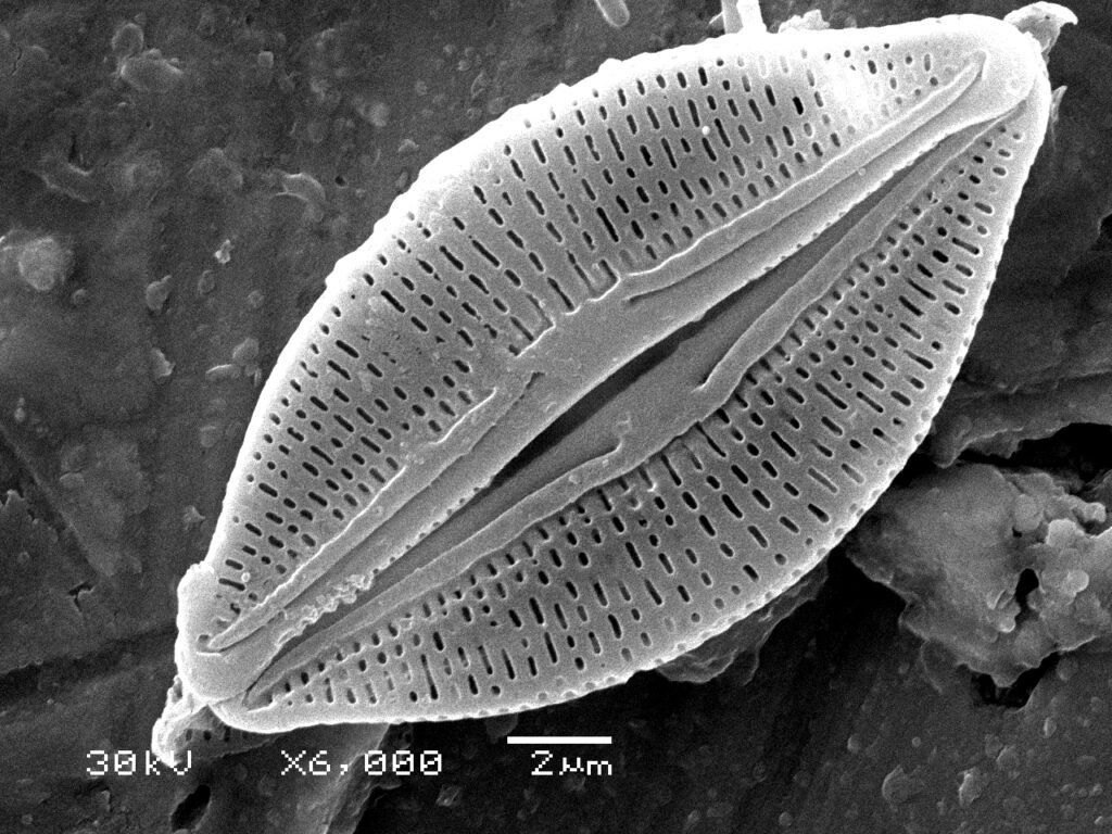
Diatoms
Diatoms are single-celled algae, specifically microalgae, that live in the Earth’s oceans, waterways, and soils. They are autotrophic organisms that live by photosynthesis in the photic zone – in the first 200 meters of the water column.

Blepharisma Ciliate
Blepharisma ciliate protozoan, coloured scanning electron micrograph (SEM). These tiny single-celled organisms are found in freshwater and marine habitats. It feeds on bacteria and decaying organic matter, which it filters through membranelles in its primitive mouth, known as a buccal cavity (upper centre). The membranelles, rows of fused cilia (short microscopic hairs), are also used for locomotion. Magnification: x500 when printed at 10 centimetres wide.

Synchatacta Tranceluecnt Predactory Rotiber
The predatory Synchaeta Rotifer is among the smallest of animals. Rotifers have a complete digestive tract with a feeding apparatus (mastax) and a muscular chamber equipped with jaw-like trophi. Phase contrast light micrograph. Magnification: x16 when printed at 10 centimetres wide.

Prootozoan Amoba
The Protozoan Amoeba proteus moves by means of its pseudopodia or false feet. Differential interference contrast light micrograph. This freshwater single-celled organism feeds on bacteria and smaller protozoa. Its uses pseudopodia (limb-like extensions) to engulf food and for locomotion. Magnification: x200 when printed at 10 centimetres wide.

Bdelloid Rotifer, Rotaria Rotatoria.
Rotaria neptunia has an unusually long foot. The total length of some individuals may exceed 1 mm. B: Detail from a plate in Baker (1764) showing some of the rotifers he had observed. He described the rotifers in Fig. 1 as having "Tails enormously long". They were probably Rotaria neptunia, the first time this species was noted and drawn.
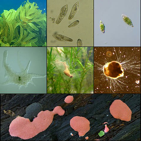
What are protists and why are they important to our ecosystem?
A Protist is a eukaryotic, unicellular organism that is aquatic, microscopic, and has a nucleus. Protists are not considered plants, animals, or fungi. They are highly diverse and considered highly organized. A Protist is highly organized due to being a eukaryote. A eukaryote has a cell that includes a nucleus and other organelles. Examples of Protist include Euglena, Amoebas, Paramecium, and Diatoms. These organisms all live in water and are made up of one cell with specialized organelles. A Protist can have specialized organelles, such as chloroplasts, mitochondria, endoplasmic reticulum, and lysosomes.
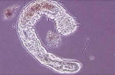
Aeolosoma (Annelida)
These transparent microannelids inhabit soils and decaying material in stagnant water, using cilia to move about. Like other annelids, Aeolosoma has a segmented body (roundworms and flatworms aren't segmented), generally consisting of about 17 segments. All but the first segment, after the head, bear sets of bristle-like structures called setae or chaetae.
The mouth is fringed with constantly moving cilia that create a vacuum cleaner effect, whisking up microscopic plants and organisms. The worms range in size from 1-2 millimeters, but often occur in long chains of immature worms, called zooids, up to 10 millimeters. These chains are produced by asexual budding, the means by which Aeolosoma reproduces.
These aquatic worms belong to the family Aeolosomatidae, which was recently put in its own class, Aphanoneura, along with the class Potamodrilidae. Some biologists, however, still classify this tiny worm in the class Oligochaeta, the same class to which the common earthworm belongs.
Currently, more than 830 species of annelids representing 27 families,12 orders, and five classes (Oligochaeta, Aphanoneura, Branchiobdellae, Acanthobdellae, and Hirudinea) are recognized as occurring in the U.S. and Canada; these include both native and introduced species.

Halteria
Halteria, a genus of microscopic planktonic ciliates that are found in many freshwater environments, can eat huge numbers of infectious chloroviruses — up to one million viruses per day — that share their aquatic habitat. Chloroviruses are known to infect microscopic green algae. Eventually, the invading chloroviruses burst their single-celled hosts like balloons, spilling carbon and other life-sustaining elements into the open water.
That carbon, which might have gone to predators of the tiny creatures, instead gets vacuumed up by other microorganisms.“That’s really just keeping carbon down in this sort of microbial soup layer, keeping grazers from taking energy up the food chain,” said lead author Dr. John DeLong, a researcher at the University of Nebraska-Lincoln.
“But if ciliates are having those same viruses for dinner, then virovory could be counterbalancing the carbon recycling that the viruses are known to perpetuate.”“It’s possible that virovory is aiding and abetting carbon’s escape from the dregs of the food chain, granting it an upward mobility that viruses otherwise suppress.”“If you multiply a crude estimate of how many viruses there are, how many ciliates there are and how much water there is, it comes out to this massive amount of energy movement (up the food chain).”“If this is happening at the scale that we think it could be, it should completely change our view on global carbon cycling.”
For their study, Dr. John and his colleagues collected samples of the water from a nearby pond.Back at the lab, they corralled all of the microorganisms they could manage, regardless of the species, into drops of the water. Finally, they added generous portions of chlorovirus.
After 24 hours, they would search the drops for a sign that any species seemed to be enjoying the company of the chlorovirus — that even one species was treating the virus less like a threat than a snack. In Halteria, they found it.“At first, it was just a suggestion that there were more of them. But then they were big enough that I could actually grab some with a pipette tip, put them in a clean drop, and be able to count them,” Dr. DeLong said.The number of chloroviruses was plummeting by as much as 100-fold in just two days.The population of Halteria, with nothing to eat but the virus, was growing an average of about 15 times larger over that same timespan.Halteria deprived of the chlorovirus, meanwhile, wasn’t growing at all.To confirm that Halteria was actually consuming the virus, the authors tagged some of the chlorovirus DNA with a fluorescent green dye before introducing the virus to the ciliates.Sure enough, the ciliate equivalent of a stomach, its vacuole, was soon glowing green.It was unmistakable: the ciliates were eating the virus. And that virus was sustaining them.“I was calling up my co-authors: ‘They grew! We did it!’ I’m thrilled to be able to see something so fundamental for the first time,” Dr. DeLong said.The researchers have since identified other ciliates that, like Halteria, can thrive by dining on viruses alone.The more they uncover, the more likely it seems that virovory could be occurring in the wild.
Scientist: Dr. Alireza Kamali Dehkordi All rights reserved 2023
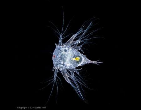
NAUPLII LARVAE
Crustaceans begin life as an egg and then go through a series of larval stages, molting several times, before reaching adulthood. The first larval stage for crabs, lobsters, shrimp, barnacles, copepods, and some other crustaceans is called a nauplius. The nauplii of different species all look alike so they are hard to tell apart. These larvae live in the water column as part of the zooplankton.They’re stealthy little creatures in that they blend in with many zooplankton. Because of this, predators have a hard time seeing them.
Nauplii look especially otherworldly and do not resemble how they will look as adult crustaceans. Like other zooplankton, they have feathery appendages to keep them from sinking to the bottom. The feathers on their heads propel their swimming. Many nauplii don’t even have mouthparts, so they don’t eat. Unlike the adult, the nauplius is a Cyclops: it only has one eye. The nauplii change form several times, adding appendages and segments as they grow with each molt. Many ocean explorers tow for Plankton. There’s a lot we can learn from plankton and the nauplii is no exception. Sometimes nauplii of various species have barnacles that are easy to identify as they have ‘horns’ that distinguish them. Nauplii are an important food source for fish and predatory invertebrates. Their health is essential to other marine life.